Home

פרח עירוני דולר קול nucleolus light microscope וויסקי מהירות צנרת

The cell. 4. Nucleus. Nucleolus. Atlas of plant and animal histology.

Nucleolar dynamics and interactions with nucleoplasm in living cells | eLife

cell nucleus Dr.Jastrow's EM-Atlas

Cell Nucleus - function, structure, and under a microscope - Rs' Science

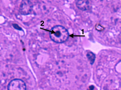
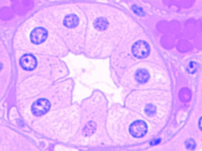
Human Hepatocyte. Nucleolus Stock Photo - Image of nucleolus, microscopic:  242799270
Human Hepatocyte. Nucleolus Stock Photo - Image of nucleolus, microscopic: 242799270

Basic Histology -- Nucleoli

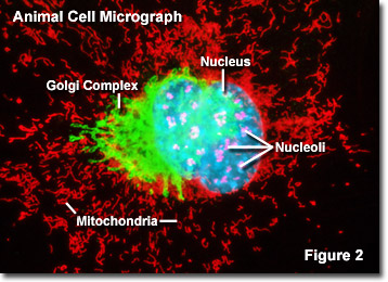
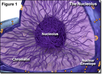
Molecular Expressions Cell Biology: The Nucleolus

To be or not to be in the nucleolus | Nature Cell Biology

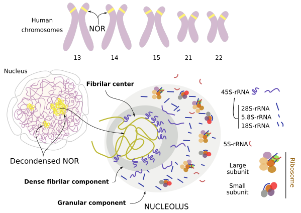
Nucleolus - Wikipedia

Nucleolus - an overview | ScienceDirect Topics

Carbon Dots for Single-Molecule Imaging of the Nucleolus | ACS Applied Nano  Materials
Carbon Dots for Single-Molecule Imaging of the Nucleolus | ACS Applied Nano Materials

Photograph | Hepatocyte, Nucleolus, Amitosis, LM | Science Source Images

Cell Lab
Cell Lab

Question Video: Identifying the Parts of the Nucleus in an Electron  Micrograph | Nagwa
Question Video: Identifying the Parts of the Nucleus in an Electron Micrograph | Nagwa

Cell Lab
Cell Lab

Biology Coach on Twitter: "Last week's #MysteryAnatomy structure was the # Nucleolus. Visible under a light #microscope, it is the largest structure  in the #nucleus. Containing #DNA & #RNA (which carry the instructions
Biology Coach on Twitter: "Last week's #MysteryAnatomy structure was the # Nucleolus. Visible under a light #microscope, it is the largest structure in the #nucleus. Containing #DNA & #RNA (which carry the instructions

Molecular Expressions Cell Biology: The Nucleolus

An electron micrograph of a barley nucleus including a nucleolus (N).... |  Download Scientific Diagram
An electron micrograph of a barley nucleus including a nucleolus (N).... | Download Scientific Diagram

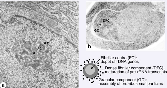
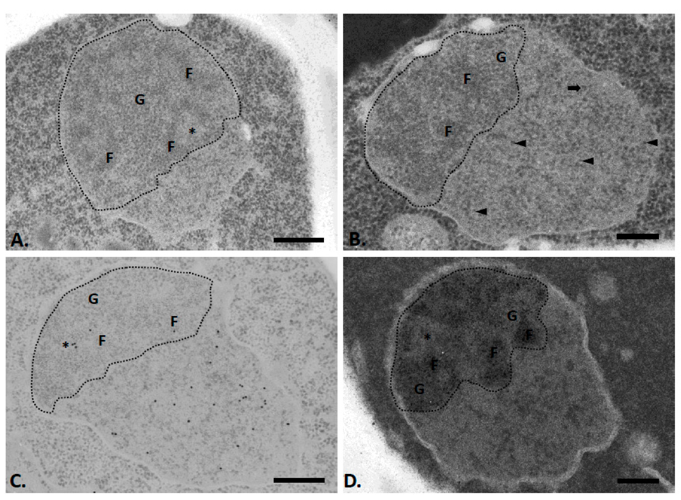
Structural and Functional Organization of Ribosomal Genes within the  Mammalian Cell Nucleolus - Massimo Derenzini, Gianandrea Pasquinelli,  Marie-Francçoise O'Donohue, Dominique Ploton, Marc Thiry, 2006
Structural and Functional Organization of Ribosomal Genes within the Mammalian Cell Nucleolus - Massimo Derenzini, Gianandrea Pasquinelli, Marie-Francçoise O'Donohue, Dominique Ploton, Marc Thiry, 2006
Nucleolus - Wikipedia

IJMS | Free Full-Text | Visualization of Chromatin in the Yeast Nucleus and  Nucleolus Using Hyperosmotic Shock
IJMS | Free Full-Text | Visualization of Chromatin in the Yeast Nucleus and Nucleolus Using Hyperosmotic Shock

Environmental cues induce a long noncoding RNA–dependent remodeling of the  nucleolus | Molecular Biology of the Cell
Environmental cues induce a long noncoding RNA–dependent remodeling of the nucleolus | Molecular Biology of the Cell

2,031 Nucleolus Images, Stock Photos & Vectors | Shutterstock

An electron micrograph of a HeLa cell demonstrates that the nucleolus... |  Download Scientific Diagram
An electron micrograph of a HeLa cell demonstrates that the nucleolus... | Download Scientific Diagram

Nucleolus. Hepatocyte stock photo. Image of histology - 93292624

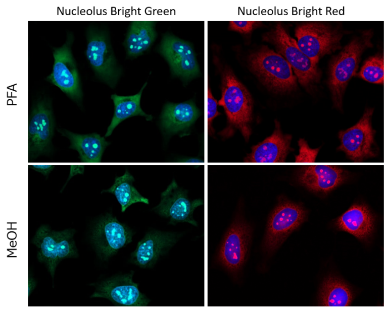
Nucleolus Fluorescent Staining Nucleolus Bright Green DOJINDO

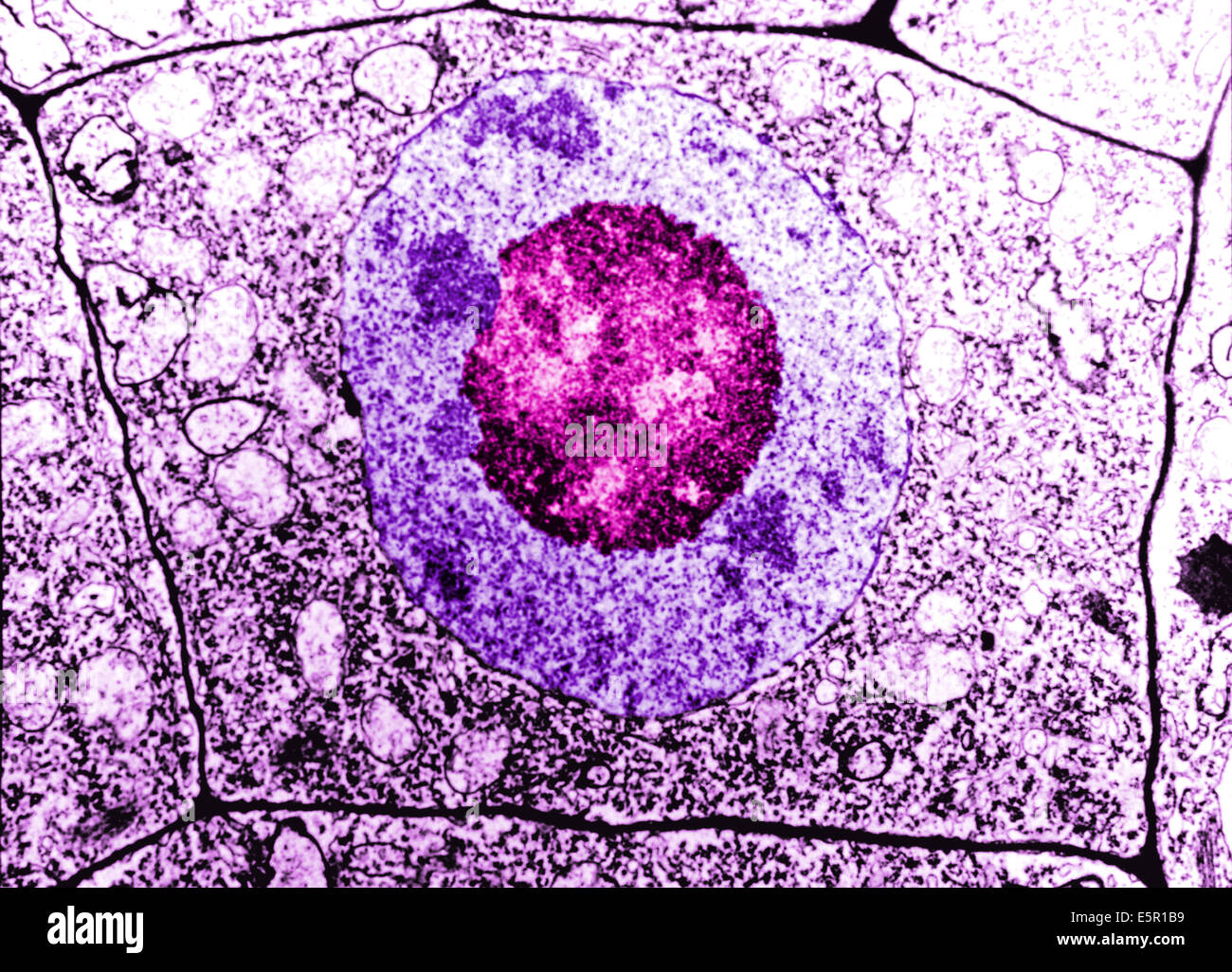
Cell nucleus tem hi-res stock photography and images - Alamy

Visualization of the Cell Using EM | Scanning electron microscope, Electron  microscope, Microscopic photography
Visualization of the Cell Using EM | Scanning electron microscope, Electron microscope, Microscopic photography